What Is Wet Macular Degeneration?
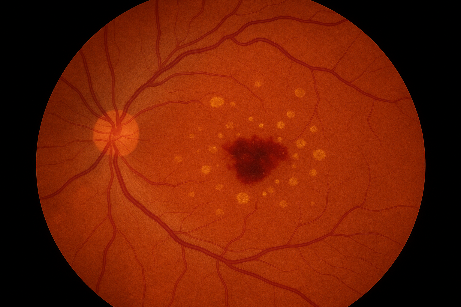
Wet Age-Related Macular Degeneration (Wet AMD) is a serious eye condition that affects the macula, the part of the retina responsible for sharp, central vision. In Wet AMD, abnormal blood vessels grow beneath the retina and leak fluid or blood, leading to swelling, damage to the macula, and sudden vision loss if not treated promptly.
How Is Wet AMD Treated?
At our clinic, Wet AMD is treated with Avastin® (bevacizumab) injections, a highly effective medication that helps reduce fluid, stop abnormal blood vessel growth, and stabilize or improve vision.
Avastin is administered directly into the eye in a quick, in-office procedure. Most patients require a series of injections spaced out over several months, with ongoing monitoring to assess the response and adjust the treatment plan as needed.
Early intervention and consistent follow-up are key to preserving vision, and we are committed to providing personalized care every step of the way.
Why Does Wet AMD Happen?
Wet AMD typically develops as an advanced form of dry macular degeneration and is most common in individuals over the age of 60. The exact cause is not fully understood, but several risk factors can contribute, including:
- Age
- Family history of AMD
- Smoking
- High blood pressure or cardiovascular disease
- Prolonged exposure to ultraviolet (UV) light
Though Wet AMD accounts for a smaller percentage of AMD cases, it causes the most severe vision loss associated with the condition.
How Do We Test for Wet AMD?
Early detection is critical, as Wet AMD can progress rapidly. At our clinic, we use advanced imaging to monitor and diagnose changes in the retina, including:
- Optical Coherence Tomography (OCT) – Provides high-resolution, cross-sectional images of the retina to detect fluid, swelling, or structural changes in the macula.
- Optos widefield retinal imaging – Offers a comprehensive view of the retina, helping us assess overall eye health and detect peripheral changes.
- OCT-Angiography (OCT-A) – A non-invasive scan that maps blood flow in the retina, making it possible to detect abnormal vessels without the need for contrast dye.
- Visual acuity testing and Amsler grid monitoring – Used to assess central vision and detect distortion or blind spots that may indicate macular changes.